Quick Answer (30 seconds)
Successful CT detector array board layout requires managing extreme signal sensitivity and high-density interconnects (HDI) within a strictly controlled thermal environment. The primary goal is to minimize noise in the analog front end (AFE) while maintaining precise geometric alignment of photodiodes.
- Material Selection: Use high-performance laminates like Rogers or Megtron 6 to minimize dielectric loss and ensure dimensional stability.
- Signal Integrity: Isolate analog signals from digital switching noise using dedicated ground planes and guard traces.
- Layout Precision: Photodiode pads must align within microns to prevent image artifacts; use non-solder mask defined (NSMD) pads for better registration where applicable.
- Thermal Management: Implement thermal vias and copper balancing to prevent warping, which causes ring artifacts in CT images.
- Cleanliness: Strict ionic cleanliness is required to prevent leakage currents in high-impedance circuits.
- Validation: Always perform impedance simulation and thermal modeling before fabrication.
When CT detector array board layout applies (and when it doesn’t)
Understanding the specific context of your design ensures that the rigorous standards of medical imaging are applied only where necessary.
This guide applies to:
- Medical CT Scanners: High-slice count (64, 128, 256+) systems requiring ultra-low noise floors and precise photodiode arrays.
- Industrial CT (NDT): Non-destructive testing equipment used for inspecting aerospace components or electronics, where resolution is critical.
- Security Baggage Scanners: Multi-energy X-ray systems that utilize similar detector array architectures for material discrimination.
- Dental CBCT: Cone Beam CT systems that require compact, high-density detector layouts.
- Replacement Detector Modules: Reverse engineering or upgrading legacy detector boards for extended equipment life.
This guide does not apply to:
- Standard MRI Coils: While medical, MRI relies on RF resonance and magnetic fields, requiring non-magnetic materials and different layout rules.
- General Consumer Electronics: Standard FR4 boards do not meet the leakage current or dimensional stability requirements of CT detectors.
- Flat Panel Detectors (DR): Digital Radiography panels use amorphous silicon TFT arrays on glass, which differs significantly from PCB-based discrete detector arrays.
- Low-Frequency Power Electronics: The noise mitigation techniques here are specific to high-impedance, low-current sensor signals, not high-power switching.
Rules & specifications
Once you confirm the application, you must adhere to strict design rules to ensure the CT detector array board functions correctly without introducing image artifacts.
| Rule | Recommended Value/Range | Why it matters | How to verify | If ignored |
|---|---|---|---|---|
| Trace Width/Space (HDI) | 3 mil / 3 mil (0.075mm) | High channel counts require dense routing to fit thousands of pixels in a small area. | Automated Optical Inspection (AOI) and DFM checks. | Short circuits or inability to route all channels, leading to dead pixels. |
| Impedance Control | 50Ω SE / 100Ω Diff (±5%) | Ensures signal integrity for high-speed ADC data transmission. | Use an Impedance Calculator during stackup design. | Signal reflections causing data corruption and image noise. |
| Material Dk/Df | Dk < 3.5, Df < 0.002 | Low dielectric absorption prevents signal loss and "ghosting" artifacts. | Review material datasheets (e.g., Rogers PCB materials). | Blurring of images and reduced contrast resolution. |
| Layer Stackup Symmetry | 100% Symmetrical | Prevents warping during reflow; critical for sensor alignment. | Check stackup construction in CAM software. | Board warpage causes sensor misalignment and "ring artifacts." |
| Analog/Digital Isolation | > 20 mil separation | Prevents digital switching noise from coupling into sensitive analog sensor lines. | Design Rule Check (DRC) with specific clearance rules. | High noise floor, rendering low-dose imaging impossible. |
| Via Aspect Ratio | Max 10:1 (Mechanical), 0.8:1 (Micro) | Ensures reliable plating in thick boards with small vias. | Cross-section analysis (microsection) after plating. | Open vias under thermal stress, leading to intermittent channel failure. |
| Surface Finish | ENEPIG or Hard Gold | Provides a flat, wire-bondable surface (if applicable) and oxidation resistance. | X-ray Fluorescence (XRF) measurement of thickness. | Poor wire bond strength or solder joint failure over time. |
| Cleanliness (Ionic) | < 1.56 µg/cm² NaCl eq. | Ionic residues cause leakage currents between high-impedance sensor traces. | ROSE testing or Ion Chromatography. | Drifting pixel values and calibration errors. |
| Thermal Vias | 0.3mm hole, filled/capped | Dissipates heat from ADCs to prevent thermal noise drift. | Thermal simulation and IR imaging of prototype. | Localized heating causes sensor drift and image inconsistencies. |
| Copper Balance | > 80% uniformity per layer | Maintains etching consistency and flatness. | CAM analysis of copper density. | Etching variation affects impedance; warping affects alignment. |
| Guard Rings | Surrounding sensitive inputs | Shunts surface leakage currents to ground, protecting the signal. | Visual inspection of layout. | Increased noise in humid environments. |
| Back-drilling | Stub length < 10 mil | Removes unused via stubs to reduce signal reflection in high-speed links. | TDR (Time Domain Reflectometry) testing. | Signal integrity issues at high data rates. |
Implementation steps
With the specifications defined, the next phase is executing the CT detector array board layout through a structured design and manufacturing process.
Define the Stackup and Materials: Start by selecting a material with a low Coefficient of Thermal Expansion (CTE) and low dielectric loss. Consult with APTPCB (APTPCB PCB Factory) early to confirm stock availability of high-performance laminates. Define a symmetrical stackup (e.g., 12-18 layers) to accommodate the high density of routing.
Place the Photodiode/Sensor Array: This is the most critical step. Place the sensor footprints based on the precise mechanical focal arc of the CT scanner. Use a mechanical CAD (MCAD) integration to ensure the coordinates are exact. Lock these components immediately to prevent accidental movement.
Route the Analog Front End (AFE): Route the traces from the photodiodes to the Analog-to-Digital Converters (ADCs). These traces must be as short as possible and matched in length to ensure phase consistency. Use guard traces or ground pours to shield these lines from external noise.
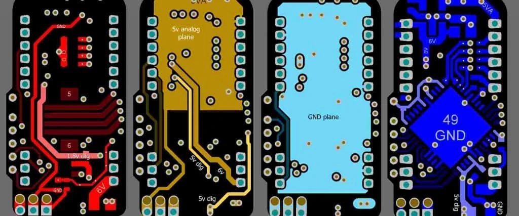
Implement Power Distribution: Design the power planes to provide clean, stable power to the ADCs. Use multiple decoupling capacitors placed close to the power pins. Separate the analog power (AVDD) from the digital power (DVDD) using ferrite beads or separate regulator outputs.
Route Digital Data Lines: Route the high-speed digital outputs from the ADCs to the Data Acquisition System (DAS) interface. Maintain strict impedance control (usually 100Ω differential). Avoid crossing splits in the ground plane, which creates return path discontinuities and EMI issues.
Apply Shielding and Grounding: Create a solid ground reference plane immediately adjacent to the signal layers. Stitch the ground planes together with a dense array of vias to minimize ground impedance. Ensure the chassis ground is connected correctly at the mounting holes but isolated from the signal ground if required by the system architecture.
Perform DFM and DFA Checks: Run a comprehensive Design for Manufacturing (DFM) check. Verify minimum trace widths, annular rings, and mask clearances. Check for "acid traps" (acute angles) in the layout. Refer to DFM Guidelines to ensure the board can be manufactured with high yield.
Generate Manufacturing Files: Output ODB++ or Gerber X2 files. Include a detailed fabrication drawing specifying the material, impedance requirements, and tolerance classes (e.g., IPC Class 3 for medical).
Failure modes & troubleshooting
Even with a rigorous design process, issues can arise during testing; systematic troubleshooting of the CT detector array board is essential to identify root causes.
Symptom: Ring Artifacts in Image
- Cause: Non-uniform sensitivity or gain across the detector channels, often due to board warpage or inconsistent trace impedance.
- Check: Measure the flatness of the board. Verify trace widths on the outer channels versus inner channels.
- Fix: Recalibrate the detector gain map. If physical warping is present, redesign the stackup for better copper balance.
- Prevention: Use high-Tg materials and ensure 100% symmetrical stackup.
Symptom: High Noise Floor (Grainy Image)
- Cause: Coupling of digital noise into analog signals or poor grounding.
- Check: Use a spectrum analyzer to look for switching frequencies on the analog supply. Check for ground loops.
- Check: Verify that analog and digital return paths do not overlap.
- Fix: Add shielding cans or improve decoupling. Cut ground loops if possible.
- Prevention: Strict separation of analog and digital sections in the layout phase.
Symptom: Channel Crosstalk (Ghosting)
- Cause: Signal traces routed too closely together without adequate isolation.
- Check: Inject a signal into one channel and measure the output on adjacent channels.
- Fix: It is difficult to fix on a finished board. Software correction may help.
- Prevention: Follow the "3W rule" (spacing = 3x trace width) for sensitive signals. Use ground guard traces.
Symptom: Thermal Drift (Signal changes over time)
- Cause: Components heating up and changing characteristics, or PCB expansion shifting sensor alignment.
- Check: Monitor board temperature with a thermal camera during operation.
- Fix: Improve airflow or add heatsinks to hot components.
- Prevention: Design adequate thermal vias and copper planes for heat dissipation.
Symptom: Intermittent Open Circuits
- Cause: Micro-cracks in vias or solder joints due to thermal cycling (CT scanners spin fast and generate heat).
- Check: Perform thermal cycling tests. Use X-ray inspection on BGA/LGA components.
- Fix: Reflow or replace the component. If via failure, the board is scrap.
- Prevention: Limit via aspect ratio. Use corner bonding or underfill for large BGAs.
Symptom: Leakage Current Errors
- Cause: Ionic contamination on the board surface bridging high-impedance traces.
- Check: Perform localized cleanliness testing. Look for flux residue.
- Fix: Clean the board using an ultrasonic bath with specialized saponifiers.
- Prevention: Specify strict cleanliness standards (e.g., < 1.0 µg/cm² NaCl) in fabrication notes.
FAQ
Troubleshooting often leads to specific questions about manufacturing capabilities and design trade-offs for CT detector array board design.
Q: What is the best PCB material for CT detector boards? A: High-performance materials like Rogers 4000 series or Panasonic Megtron 6 are preferred. They offer low dielectric loss and excellent dimensional stability, which is crucial for maintaining sensor alignment under thermal stress.
Q: Why is impedance control critical for detector boards? A: Impedance mismatches cause signal reflections, which degrade the integrity of the high-speed digital data sent from the ADCs to the image processor. This results in data errors and image artifacts.
Q: Can I use standard FR4 for a prototype? A: It is not recommended. FR4 has higher dielectric loss and less consistent mechanical properties than required. Data collected from an FR4 prototype may not accurately represent the performance of the final medical device.
Q: How do I handle the high density of connections? A: Use High-Density Interconnect (HDI) technology, including blind and buried vias. This allows you to route signals on inner layers without consuming space on the surface, enabling tighter component placement.
Q: What is the typical lead time for these boards? A: Due to the complexity (high layer count, special materials, Class 3 requirements), lead times are typically longer than standard boards. Expect 3-4 weeks for fabrication, plus additional time for assembly and testing.
Q: How does APTPCB ensure the cleanliness of these boards? A: We utilize advanced cleaning lines and perform Ionic Contamination Testing (ROSE test) to ensure residues are below medical-grade limits. This prevents leakage currents that can corrupt sensor data.
Q: Is underfill required for the ADCs or ASICs? A: Often, yes. The high G-forces generated by the rotating gantry of a CT scanner put mechanical stress on solder joints. Underfill provides mechanical reinforcement to prevent fatigue failures.
Q: What surface finish is best for wire bonding photodiodes? A: ENEPIG (Electroless Nickel Electroless Palladium Immersion Gold) or Soft Gold is recommended. These finishes provide a pure gold surface that is ideal for reliable wire bonding.
Q: How do I prevent "starving" the thermal pads during reflow? A: Avoid placing large open vias directly in thermal pads unless they are filled and capped. Open vias can wick solder away from the joint, leading to poor thermal connection and component overheating.
Q: Do I need to simulate the board before manufacturing? A: Yes. Signal integrity (SI) and power integrity (PI) simulations are highly recommended. They help identify potential crosstalk and power delivery issues before physical prototypes are built, saving time and cost.
Glossary (key terms)
Familiarity with these terms is essential for effective communication regarding CT detector array board assembly and fabrication.
| Term | Definition | Context in CT Detectors |
|---|---|---|
| AFE (Analog Front End) | The circuitry that interfaces directly with the sensors to condition the signal. | The most noise-sensitive section of the board; requires careful layout. |
| Photodiode | A semiconductor device that converts light (from the scintillator) into electrical current. | The primary sensor element; requires precise mechanical alignment. |
| Scintillator | A material that converts X-ray photons into visible light. | Mounted on top of the photodiodes; alignment with the PCB is critical. |
| HDI (High-Density Interconnect) | PCB technology using microvias, blind/buried vias, and fine lines. | Essential for routing thousands of channels in a compact detector arc. |
| Crosstalk | Unwanted signal transfer between communication channels. | Causes "ghosting" or blurring between adjacent pixels in the image. |
| CTE (Coefficient of Thermal Expansion) | The rate at which a material expands with temperature. | Mismatch between PCB and components causes stress and warping. |
| Dark Current | The residual current flowing in a photodiode when no light is present. | Must be minimized and calibrated out; leakage on PCB increases this. |
| IPC Class 3 | The highest standard for PCB reliability (High Performance Electronic Products). | Required for medical life-support and critical diagnostic equipment. |
| Anti-Scatter Grid (ASG) | A grid placed over the detector to block scattered X-rays. | The PCB layout must align perfectly with the mechanical ASG structure. |
| DAS (Data Acquisition System) | The system that collects digital data from the detector modules. | The destination for the high-speed signals routed from the detector board. |
| Microvia | A laser-drilled via with a diameter typically less than 150 microns. | Used to connect surface layers to inner layers in HDI designs. |
| Anisotropic Conductive Film (ACF) | A tape used to connect flex circuits or glass panels to PCBs. | Sometimes used to connect the sensor array to the main readout board. |
Conclusion
Designing a CT detector array board layout is a balancing act between electrical performance, mechanical precision, and thermal stability. Every trace width, via placement, and material choice directly impacts the diagnostic quality of the final image. By adhering to strict design rules—such as impedance control, analog isolation, and rigorous cleanliness—you can eliminate artifacts and ensure long-term reliability.
Whether you are prototyping a new multi-slice scanner or maintaining legacy medical equipment, APTPCB provides the specialized manufacturing capabilities required for medical-grade electronics. From high-frequency material sourcing to IPC Class 3 fabrication, we ensure your design meets the highest standards of safety and performance.
Ready to validate your medical PCB design? Request a PCB Quote today or explore our PCB Manufacturing Capabilities to see how we can support your next imaging project.